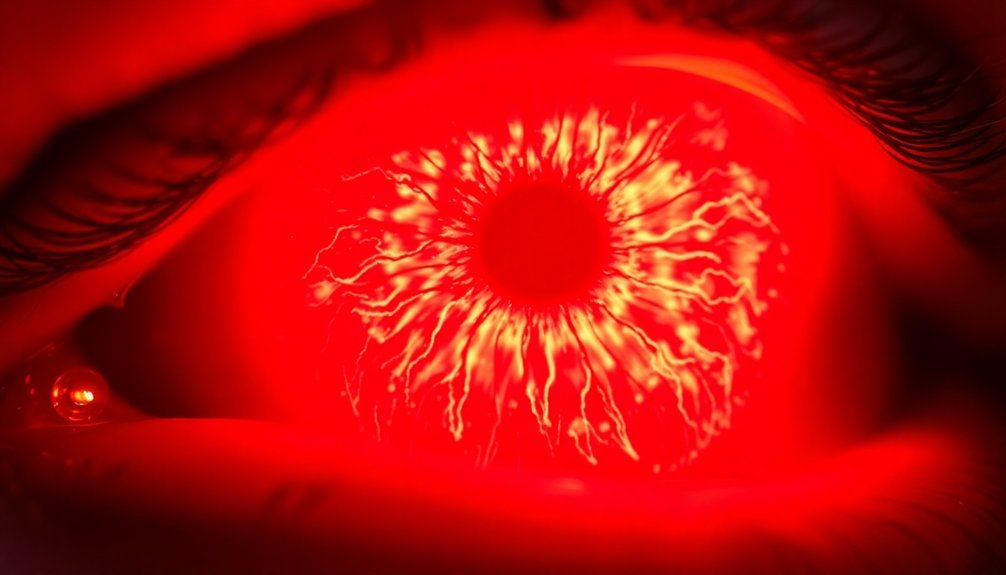
LED therapy helps restore your corneal tissue through several key mechanisms. When specific wavelengths of LED light hit your cornea, they activate the mitochondria in your cells, boosting ATP production and cellular energy. This increased energy helps your cells repair and regenerate faster. The light also triggers important growth factors like EGF and HGF, which promote cell migration and proliferation while reducing scar formation. Blue LED light (405-450nm) can even help form stable, tissue-like structures, while red light activates healing pathways. Understanding these biological responses can help you maximize your treatment's effectiveness.
Understanding Corneal Tissue Repair Mechanisms

The cornea's remarkable repair process kicks off through three main mechanisms: migration, proliferation, and remodeling.
When your cornea sustains damage, epithelial cells immediately begin migrating toward the injured site, while limbal stem cells produce specialized cells called transient amplifying cells that move to repair the damage. Growth factors in tears are essential for maintaining normal ocular surface function during this process.
During proliferation, these transient amplifying cells multiply and transform into new corneal epithelial cells to fill the wound. Your basal cells divide through mitosis, moving both inward and upward to close the gap, while tight junctions reform to restore the cornea's protective barrier function.
The final phase involves differentiation and remodeling, where the extracellular matrix undergoes restructuring to maintain the stroma's integrity. Growth factors play vital roles throughout this process – EGF, HB-EGF, and TGF-α orchestrate the repair, while substances like HDAC6 promote cell migration.
Your limbal stem cells are particularly important, as they're responsible for continuous corneal regeneration throughout your life. They're supported by mesenchymal stem cells, which secrete healing factors like HGF to reduce inflammation and promote recovery.
LED Light Effects On Eyes
While understanding corneal repair mechanisms helps guide treatment approaches, it's equally important to take into account potential risks when using LED therapy for eye conditions.
The effects of LED light exposure on your eyes can be both immediate and long-lasting, requiring careful consideration before starting any treatment protocol. LEDs convert up to 90% less energy to heat compared to traditional lighting, which helps reduce thermal damage during treatments.
You'll need to be aware that LED exposure can cause irreversible retinal damage and accelerate tissue aging. When using LED therapy for corneal treatment, protect yourself from these potential hazards:
- Wear appropriate eye protection specifically designed for LED therapy
- Follow strict exposure time limits as prescribed by your healthcare provider
- Use only FDA-cleared devices for corneal treatment
- Maintain proper distance between the light source and your eye
- Avoid treatment during evening hours to prevent circadian rhythm disruption
Even short-term exposure can lead to visual fatigue, headaches, and blurred vision. The invisible flicker in LEDs may also affect your visual performance during treatment sessions.
If you're considering LED therapy for corneal tissue repair, you'll need to carefully weigh these risks against potential benefits and work closely with your eye care professional to establish a safe treatment protocol.
Cellular Response To Light Therapy

When you expose corneal tissue to LED therapy, mitochondrial activity dramatically increases, leading to enhanced ATP production and cellular energy levels.
You'll find that this boost in cellular energy triggers specific growth factor pathways, which help regulate cell repair and regeneration processes. Studies show that wavelengths above 660nm are particularly effective at stimulating these healing responses.
The light therapy also sets off powerful anti-inflammatory mechanisms in your corneal cells, reducing harmful inflammatory cytokines while promoting tissue healing and repair.
Mitochondrial Energy Production Boost
Light therapy's remarkable ability to boost mitochondrial energy production lies at the heart of its therapeutic effects on corneal tissue. When specific wavelengths of light reach your cells, they activate cytochrome oxidase IV (COX IV), a vital enzyme in your mitochondria that's responsible for cellular energy production.
This activation triggers several beneficial responses in your corneal cells:
- Increased ATP production for enhanced cellular energy
- Improved resistance to toxic substances and oxidative stress
- Enhanced cell survival and reduced apoptosis rates
- Faster cellular proliferation and recovery
- Greater protection against environmental damage
You'll find that this boost in mitochondrial function is particularly effective for treating corneal conditions. When your corneal endothelial cells receive red light therapy, they respond by increasing their energy production and improving their survival mechanisms. The carefully controlled pulsed blue light exposure helps maintain optimal tissue response while preventing potential toxicity.
This enhanced energy state helps your cells better resist damage from various sources, including toxic substances like sodium azide and cobalt chloride.
The process, known as photobiomodulation, works through low-energy light therapy that specifically targets mitochondrial enzymes. It's a non-invasive approach that's proven especially effective in treating conditions like dry eye disease, where it helps reduce inflammation and improve corneal surface irregularities.
Growth Factor Pathway Activation
Building on cellular energy enhancement, LED therapy's effects extend deeply into your body's growth factor pathways. When red LED light reaches your corneal tissue, it triggers a cascade of cellular responses, particularly through the epidermal growth factor receptor (EGFR) pathway. This activation stimulates vital proteins like Akt and NF-κB, which drive cell proliferation and migration necessary for healing.
You'll find that LED therapy doesn't work alone – it enhances the effectiveness of natural growth factors in your cornea. The treatment amplifies EGF's healing properties while promoting the expression of TGF-β, creating a powerful combination for tissue repair. It's especially effective at mobilizing limbal stem cells, which are essential for corneal regeneration.
The therapy's influence on growth factors goes beyond basic repair. When you receive red LED treatment, it activates TRPV1 channels, reducing scar formation while promoting healthy cell growth. It also controls inflammation through COX-2 modulation and enhances blood vessel formation via VEGF expression. This coordinated response helps safeguard your corneal tissue heals properly while maintaining its transparency and function.
Anti-Inflammatory Response Mechanisms
Through advanced cellular mechanisms, LED therapy triggers a powerful anti-inflammatory response in corneal tissue. When specific wavelengths of red and near-infrared light penetrate the cornea, they activate cytochrome c oxidase in the mitochondria, leading to increased ATP production and enhanced cellular survival. This activation directly reduces inflammation by suppressing key inflammatory markers.
The anti-inflammatory effects of LED therapy are particularly evident in treating dry eye disease, where 740 nm light substantially decreases inflammatory cytokines. You'll see reduced levels of:
- IL-1β cytokines
- IL-6α markers
- TNF-α inflammation factors
- VEGF-C/D expression
- Corneal lymphatic diameter
Your corneal tissue responds to LED therapy through multiple pathways. The infrared light penetrates deeper into the tissue, where it accelerates healing and reduces inflammation.
This process works in concert with the activation of mitochondrial protective mechanisms, which shield corneal endothelial cells from mechanical damage.
Additionally, the therapy's effect on lymphatic vessels helps control inflammation, as blocking VEGF-C results in decreased lymphatic vessel size and area, further supporting the cornea's healing process.
Growth Factors During Healing
Growth factors step up to play vital roles during corneal healing, orchestrating a complex series of cellular responses that promote tissue repair. EGF and HB-EGF are particularly active, stimulating cell growth and mobility while working in an autocrine manner on epithelial cells.
When your cornea is healing, BTC accelerates the process by promoting limbal stem cell proliferation through ERK1/2 phosphorylation.
You can think of NGF as your cornea's nerve regeneration specialist, while TGF-α helps regulate overall cell growth patterns. These growth factors don't work alone – they're supported by substance P and BDNF, which contribute substantially to the wound healing process.
You'll see enhanced expression of specific keratins (1, 10, and 17) during light-treated wound healing, indicating improved epithelialization.
What's particularly interesting is how blue LED light therapy can influence these growth factors' activity. It's not just about the growth factors themselves – it's about how they interact with light therapy to create an ideal healing environment.
The combination of proper growth factor signaling and light therapy can markedly speed up your cornea's natural healing process.
Wavelength Selection For Corneal Treatment
Selecting the right wavelength proves essential for effective corneal treatment, with low-energy blue light emerging as the primary choice. When treating damaged corneal tissue, you'll find that the 405-450 nm spectrum of blue light effectively activates specialized biomaterials designed to reshape and thicken the cornea.
The treatment's success relies on pulsed irradiation, alternating between 2.5 seconds on and off, which enhances hydrogel formation and oxygen recovery. You're leveraging a carefully engineered process where blue light triggers the assembly of short peptides and glycosaminoglycans into a stable, tissue-like structure.
Key characteristics that make blue light ideal for corneal treatment include:
- Lower penetration depth compared to red light wavelengths
- Specific activation of biomaterial assembly
- Safe interaction with corneal tissue
- Ideal hydrogel formation through pulsed delivery
- Minimal cytotoxic effects due to low-energy application
You'll notice this approach differs substantially from other LED therapies, such as red light used for deeper tissue penetration. The careful wavelength selection guarantees you're targeting the precise depth needed for corneal repair while maintaining safety and effectiveness throughout the treatment process.
Light Therapy Safety Protocols
You'll need to carefully select wavelengths between 630-850nm when treating corneal tissue, as these ranges have demonstrated ideal therapeutic effects while maintaining safety standards.
Your device's calibration must be verified before each treatment session, ensuring consistent power output and beam uniformity across the treatment area.
Treatment duration shouldn't exceed 3-5 minutes per session during initial treatments, with adjustments made based on your patient's response and corneal tissue sensitivity.
Wavelength Selection Guidelines
Wavelength selection's critical role in LED therapy demands careful attention to safety protocols and specific therapeutic targets. When treating corneal tissue, you'll need to understand that different wavelengths penetrate to varying depths and serve distinct therapeutic purposes.
Blue light around 450-495 nm works effectively for corneal repair biomaterials, while red light (630-700 nm) and near-infrared light target different tissue layers.
For the best corneal tissue restoration, you'll want to think about these essential wavelength selection guidelines:
- Always verify FDA clearance for your chosen device and wavelength combination
- Select blue light specifically for corneal repair biomaterials activation
- Combine red and NIR wavelengths when deeper tissue penetration is needed
- Use protective eyewear designed for your specific wavelength range
- Follow manufacturer-specified exposure times and power settings
You must prioritize safety while maximizing therapeutic benefits. Professional advice is vital, especially when dealing with corneal applications.
Remember that incorrect wavelength selection can compromise both safety and treatment efficacy. When using LED therapy for corneal tissue restoration, you'll need to strictly adhere to established protocols and guarantee proper eye protection throughout each session.
Device Calibration Standards
Proper device calibration stands as the cornerstone of safe and effective LED corneal therapy. You'll need to guarantee your device maintains consistent light intensity and frequency through regular calibration checks according to the manufacturer's specifications.
When you're treating sensitive corneal tissue, even minor calibration errors can lead to reduced effectiveness or potential adverse effects.
To maintain device integrity, you should follow a strict calibration schedule and keep detailed records of each calibration session. You'll want to verify that your device meets all safety standards and regulatory requirements before each treatment session.
Remember that proper calibration isn't just about effectiveness – it's vital for protecting your patient's delicate eye tissue.
If you're using a new device, make sure it comes with proper certification from reputable organizations and complies with relevant safety standards. You'll need to perform quality control checks regularly and document any deviations from expected performance.
When in doubt about calibration procedures, don't hesitate to contact the manufacturer or consult with technical specialists. The success of corneal tissue restoration heavily depends on precise light delivery, making proper calibration an non-negotiable aspect of your treatment protocol.
Treatment Duration Parameters
Three key parameters govern safe LED therapy for corneal tissue: pulse timing, session frequency, and exposure duration. For the best results, you'll need to follow specific timing protocols, especially when using blue light for corneal repair.
Research shows that alternating 2.5-second pulses of light with 2.5-second rest intervals produces the most favorable outcomes while maintaining tissue safety.
When it comes to treatment frequency, you should space your sessions every other day over several weeks. This spacing allows proper tissue recovery and prevents overexposure. During each session, you'll need to wear specialized protective goggles to shield your eyes from direct light exposure.
To guarantee safe and effective treatment, follow these essential guidelines:
- Use only FDA-cleared devices with proven safety features
- Maintain low-intensity light settings to prevent heat damage
- Keep treatment sessions monitored by qualified professionals
- Apply light therapy only to prescribed treatment areas
- Follow recommended exposure times without exceeding them
Remember that different light wavelengths serve different purposes – blue light works well for corneal repair biomaterials, while red LED therapy has its own specific applications. Your healthcare provider will determine the most appropriate duration parameters based on your specific condition and treatment goals.
Signaling Pathways In Healing

Molecular signaling cascades kick off a complex interplay of pathways during LED-based corneal healing. You'll find that EGFR signaling works as a primary driver, triggering downstream effects that promote tissue repair.
This process gets enhanced when specific wavelengths of LED light activate Rho-GTPase pathways, which boost cell migration without affecting proliferation rates.
When you're looking at the inflammatory response, you'll notice that Sirt6 plays a vital role in regulating pro-inflammatory cytokines like IL-1β and TNFα. Without proper Sirt6 function, you'll see excessive inflammation and delayed healing.
The MyD88 pathway also steps in, controlling chemokine expression that affects corneal thickness and clarity.
Different wavelengths trigger distinct healing mechanisms. Blue light (470 nm) enhances epithelialization, while green light (518 nm) specifically targets keratinocyte migration. Red light (638 nm) works through cytokine modulation and promotes re-epithelialization.
These wavelength-specific responses activate various molecular pathways, including mTORC2 signaling and TLR activation, which coordinate the healing process through a sophisticated network of cellular communications.
Treatment Duration And Frequency
Research-backed protocols for LED therapy in corneal treatment emphasize precise timing and delivery methods. You'll find that low-energy blue light exposure requires just 10 minutes to activate biomaterials effectively for corneal repair, with a specific dosage of 8.5 mW cm−2.
The treatment uses a pulsed light pattern of 2.5 seconds on and 2.5 seconds off, which proves more effective than continuous exposure by allowing better oxygen recovery within the hydrogel.
For ideal therapeutic outcomes, you'll need to follow these key timing parameters:
- 10-minute treatment sessions for corneal repair activation
- 5 minutes of actual light exposure when using pulsed delivery
- 2.5-second intervals for light pulsation
- 72-hour spacing between repeat treatments
- Up to 166 minutes of safe exposure time, as proven in in-vivo studies
While LED therapy protocols vary for different conditions, corneal treatment specifically demands precision in timing and delivery method. Though current research shows promising results in animal models, you should note that larger-scale studies are still needed before human clinical trials can begin.
The effectiveness of these protocols relies heavily on adhering to these research-validated durations and frequencies.
Measuring Corneal Repair Progress
You'll find several advanced imaging methods at your disposal to track corneal healing after LED therapy, including anterior segment OCT and confocal microscopy for detailed tissue assessment.
Modern biomarker analysis tools can measure key indicators like growth factors and inflammatory mediators in your corneal tissue samples, helping evaluate the repair process at a molecular level.
These assessment techniques, when combined with regular endothelial cell counts and corneal thickness measurements, give you a thorough picture of how well your cornea is responding to the LED treatment.
Imaging Assessment Methods
Advanced imaging technologies serve as the cornerstone for measuring corneal repair progress after LED therapy. You'll find that modern devices like SD-OCT and UBM provide detailed insights into healing patterns, with resolutions as fine as 2µm for precise monitoring of tissue regeneration.
These tools let you track changes in corneal thickness, evaluate epithelial conditions, and assess the effectiveness of your LED treatment protocols.
When evaluating treatment outcomes, you'll want to focus on these key imaging approaches:
- Scanning slit elevation evaluation for thorough corneal curvature assessment
- SD-OCT for high-resolution visualization of epithelial and Bowman's layer changes
- Scheimpflug-based imaging to measure biomechanical strength improvements
- UBM for detailed imaging of corneal wounds and repair
- Three-dimensional mapping to track overall healing progress
You can compare your scans against normative data to identify improvements, paying special attention to numerical overlays that indicate changes in corneal thickness and topography. For the most accurate assessment, you should analyze statistics boxes showing SimK values and zone irregularities, while maintaining consistent dioptric scales to guarantee reliable progress tracking over multiple treatment sessions.
Biomarker Analysis Tools
Beyond imaging techniques, biomarker analysis tools provide molecular and biochemical insights into corneal repair progress. You'll find that spring constant measurements through phase contrast imaging and specular microscopy can assess endothelial cell quality, giving you valuable information about long-term healing prospects.
When you're tracking corneal repair, you can rely on multiple biomarker validation methods. RT-qPCR helps you confirm gene expression patterns, while machine learning algorithms identify key markers from sequencing data.
You'll get the most accurate results by combining these with orthogonal validation approaches that connect biomarkers to clinical outcomes.
You can monitor cellular behavior and protein expression levels to gauge tissue health and regeneration potential. The Ocular Response Analyzer (ORA) measures corneal hysteresis and resistance factors, while Brillouin microscopy lets you assess corneal stiffness non-invasively. These tools work together with biochemical marker analysis to give you a complete picture of repair progress.
For precise treatment monitoring, you'll benefit from tracking specific biomarkers like apolipoprotein E (APOE). These markers don't just help you assess current healing status – they also enable early detection of complications and guide necessary therapeutic adjustments.
Combined Therapeutic Approaches
Through the integration of multiple light-based treatments, combined therapeutic approaches have emerged as a promising frontier in corneal tissue restoration. You'll find that combining photobiomodulation (PBM) therapy with blue light-activated biomaterials creates a synergistic effect that enhances healing outcomes.
These approaches work together to stimulate cellular metabolism, reduce inflammation, and promote tissue regeneration.
Key benefits of combined light therapies include:
- Increased ATP production and reduced oxidative stress
- Enhanced collagen production through red light stimulation
- Improved hydrogel stability with pulsed blue light delivery
- Better oxygen recovery within treated tissues
- Stronger anti-inflammatory response through multiwavelength exposure
When you're considering treatment options, you'll notice that devices like the Valeda Light Delivery System utilize multiple wavelengths (670-830 nm) to achieve ideal results.
The combination of PBM and LED therapy isn't just more effective – it's also showing promise as an alternative to corneal transplantation. By using pulsed light delivery techniques instead of continuous exposure, you're able to maintain better oxygen levels in the treated tissue while reducing oxidative stress, leading to more successful outcomes in corneal restoration procedures.
Frequently Asked Questions
Can LED Therapy Help With Corneal Scarring From Chemical Burns?
Yes, you'll find LED therapy helpful for chemical burn scars. Blue-light activated biomaterials can reshape and thicken your damaged corneal tissue, while forming a healing hydrogel that mimics natural corneal properties.
Does Wearing Contact Lenses Affect LED Therapy Effectiveness?
While there's no direct research on how contact lenses affect LED therapy, you'll want to consult your eye doctor as lenses might interfere with light penetration. It's best to remove them during treatment.
How Soon After LASIK Surgery Can LED Therapy Begin?
You'll need to consult your eye surgeon for specific timing, as there's no established protocol for LED therapy after LASIK. It's essential to allow proper initial healing before starting any additional treatments.
Can Children Undergo LED Therapy for Corneal Conditions?
You'll need to be cautious with LED therapy for children's corneal conditions. There's limited research on pediatric applications, so you should only proceed under strict medical supervision and after thorough evaluation by an eye specialist.
Does Eye Color Influence the Effectiveness of LED Therapy Treatment?
Your eye color doesn't substantially impact LED therapy's effectiveness, but other factors like age, lens clarity, and macular pigment density play more important roles in how well your eyes respond to the treatment.
In Summary
You'll find LED therapy's effectiveness in corneal tissue restoration stems from specific wavelengths triggering cellular repair mechanisms. When you're using the right light frequencies, they'll stimulate mitochondrial function and boost growth factors essential for healing. You can enhance your treatment outcomes by following proper protocols for duration and combining LED therapy with other proven approaches. Regular monitoring will help track your corneal repair progress.
Leave a Reply