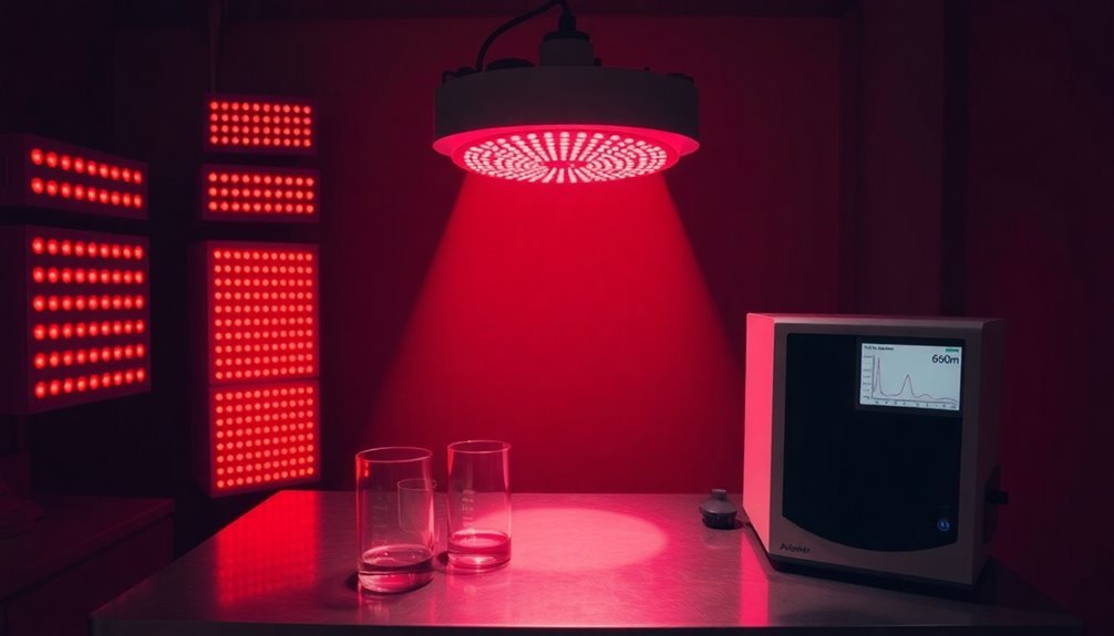
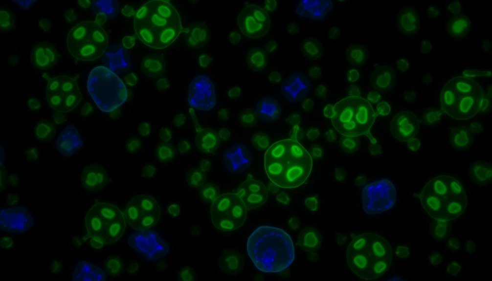
Research-backed mitochondrial recovery protocols combine multiple science-based approaches for superior results. You'll want to start with red light therapy using specific wavelengths (630-940nm) within the first 60 minutes post-stress, followed by targeted high-intensity exercise sessions to boost mitochondrial protein synthesis. Implement strategic fasting windows, like 16:8 or 5:2 protocols, to activate key genes for mitochondrial biogenesis. Include regular autophagy checks and use precise timing for both upper and lower body training protocols. Monitor progress through advanced imaging and spectroscopic techniques. These proven strategies represent just the beginning of your mitochondrial optimization journey.
Red Light Wavelength Selection

The selection of appropriate red light wavelengths plays a crucial role in mitochondrial therapy. You'll want to focus on wavelengths between 630-1000 nm, as this range provides maximal penetration and triggers the strongest mitochondrial response. This therapeutic approach is non-invasive and drug-free, making it an excellent option for long-term treatment protocols.
For targeted mitochondrial function and ATP production enhancement, you should prioritize 630 nm and 670 nm wavelengths.
When you're aiming for deeper tissue penetration and specific electron transport stimulation, opt for 850 nm and 940 nm wavelengths. These frequencies are particularly effective at activating cytochrome c oxidase and boosting your electron transport system capacity. You'll achieve better results using continuous light rather than pulsed light when targeting cellular respiration.
To maximize therapeutic benefits, you'll need to match specific wavelengths to your treatment goals. For instance, if you're treating age-related vision issues, deep red light wavelengths have shown promising results.
Different wavelengths penetrate skin to varying depths – from 595 nm to 800 nm – so you'll want to select the appropriate wavelength based on your target tissue depth. Remember, the key is to choose wavelengths that specifically target mitochondrial complexes for maximal therapeutic response.
Strategic Treatment Timing
You'll need to carefully assess mitochondrial recovery using specific markers like citrate synthase and sirtuin 3 to track treatment progress effectively.
Your ideal treatment window occurs within the first 60 minutes, when complete recovery of mitochondrial respiration and ATP levels can be achieved through targeted metabolic protocols.
To maximize peak performance timing, you should implement shortened protocols like Mito6 while monitoring the initial metabolic rate increases across repeated tests to guarantee best recovery outcomes.
Regular monitoring of GTPase activity levels through fusion and fission cycles helps determine the effectiveness of mitochondrial membrane repair protocols.
Recovery Phase Assessment Tools
Recovery phase assessment's precise timing relies on four essential diagnostic tools that researchers and clinicians use to monitor mitochondrial function: spectrophotometric assays, oxygen consumption rate measurements, nuclear magnetic resonance spectroscopy, and fluorescent probes.
Spectrophotometric assays are particularly valuable for their simplicity and reliability in measuring respiratory chain enzyme activities, requiring only small tissue samples and delivering results within three hours. ROS detection through oxidative stress markers provides critical insights into cellular damage and mitochondrial dysfunction.
The oxygen consumption rate measurements, especially using the Seahorse XF Analyzer, allow you to assess ATP production in real-time without disturbing living cells.
Nuclear magnetic resonance spectroscopy offers you thorough insights into ATP, ADP, and AMP levels while tracking TCA cycle intermediates. It's especially useful for examining mitochondrial energy metabolism in brain and skeletal muscle tissue.
For membrane potential assessment, you'll want to use fluorescent probes like Rhodamine 123, which enters the mitochondrial matrix and emits bright yellow-green fluorescence in healthy cells. These probes effectively signal changes in mitochondrial function through variations in fluorescence intensity, making them ideal for flow cytometry applications and cellular homeostasis monitoring.
Optimal Treatment Windows
Strategic timing plays a pivotal role in mitochondrial recovery protocols, with specific windows offering maximum therapeutic benefit. You'll need to focus on both immediate and long-term timing strategies to maximize your recovery outcomes.
For fasting-based interventions, you should maintain consistency with either 16:8 intermittent fasting, alternate day fasting, or the 5:2 diet protocol. These approaches activate essential genes like PGC-1alpha and Nrf2 within 24-48 hours, promoting mitochondrial biogenesis and enhanced ATP production. During fasting periods, your cells optimize energy production through increased mitochondrial splitting and fusion.
If you're undergoing mitochondrial transplantation, timing becomes even more critical. You'll need to complete the transplant within 2 hours of isolation to guarantee viability, as host cells typically engulf mitochondria within this window.
When dealing with post-injury scenarios, you must act within 24 hours, particularly for FCCP administration and mitochondrial uncoupler treatments.
Your quality control measures shouldn't be overlooked. You'll need to implement regular autophagy checks, as this process begins within hours of mitochondrial damage.
Remember to use flow cytometry and qPCR before any transplantation procedures to verify mitochondrial quality and guarantee ideal treatment outcomes.
Peak Performance Timing
Pinpointing the ideal timing for mitochondrial interventions can make or break your treatment's success. When you're targeting peak performance, you'll need to take into account both exercise intensity and sampling windows to maximize mitochondrial protein synthesis (mitoPS).
You'll achieve better results with high-intensity exercise protocols compared to moderate-intensity workouts, as they create a more potent stimulus for mitoPS. However, these effects are transient, making timing vital. The upregulation of PGC-1α and SIRT3 during exercise helps enhance mitochondrial function and reduce oxidative stress.
You'll want to carefully plan your sampling and intervention periods since arbitrary timing can lead to inconsistent or controversial results.
To optimize your performance, pay attention to your VO2 peak and ATP max measurements, as they're reliable indicators of your walking speed and overall physical capability. These markers become especially important if you're dealing with age-related mobility concerns, as they directly relate to mitochondrial efficiency.
When you're implementing protocols, you'll need validated measurement techniques, such as deuterated water testing, for accurate long-term monitoring. Remember that high-intensity exercise affects not just mitochondrial content, but also the location and activity of nuclear proteins that regulate mitochondrial function.
Targeted Body Area Protocols

To maximize mitochondrial health and recovery, protocols must be tailored to specific body regions, as different muscle groups respond uniquely to various interventions. Your upper body requires HIIT-focused exercises to enhance mitochondrial biogenesis, while your lower body benefits most from endurance training to improve mitochondrial density and function. Recent research shows that non-invasive assessment of mitochondrial oxygen metabolism can help track recovery progress in different body areas.
| Body Area | Primary Protocol |
|---|---|
| Arms & Shoulders | High-intensity intervals with resistance training |
| Legs & Glutes | Extended endurance work with dynamic movements |
| Core & Torso | Targeted strengthening with high-intensity sets |
| Back | Resistance training with mitochondrial-focused recovery |
| Full Body | Combined protocols with nutritional support |
You'll want to incorporate non-invasive measurement techniques like COMET to track your progress, particularly in accessible areas like your skin and thorax. For enhanced results, combine your targeted exercises with specific recovery strategies. Use passive acceleration techniques for lower body recovery, while implementing localized therapeutic interventions for upper body areas. Don't forget that core strengthening exercises stimulate mitochondrial biogenesis in torso muscles, making them essential for overall mitochondrial health. Support these protocols with proper nutrition and systemic exercise benefits for thorough mitochondrial optimization.
Optimal Distance And Duration
Establishing ideal distance and duration parameters is crucial for maximizing mitochondrial recovery protocols. You'll need to focus on two critical aspects: the spatial relationship between cellular components and the timing of recovery processes.
For the best distance, guarantee proper spacing between the endoplasmic reticulum (ER) and mitochondria, as this directly affects calcium uptake and oxidative metabolism. You'll want to maintain ideal proximity for efficient energy production and signaling. During respiratory conditions, mitochondrial volume increases significantly to support cellular energy demands.
Using real-time confocal microscopy, you can monitor these spatial relationships and make necessary adjustments to your protocols.
When it comes to duration, you'll need to evaluate translation timing and respiration measurements. Slower translation elongation improves mitochondrial targeting sequence recognition, while polyproline stretches in the coding sequence can create beneficial ribosome stalling.
You should adjust your measurement durations based on your specific recovery goals – shorter periods for acute responses and longer periods for tracking sustained changes.
Monitor mitochondrial volume fraction closely, as a 70% increase (as seen in chloramphenicol-treated yeast cells) can greatly improve interaction between nascent proteins and mitochondrial surfaces, enhancing overall recovery effectiveness.
Combined Healing Modalities

When you're planning mitochondrial recovery protocols, combining multiple healing approaches creates more effective results than single-method treatments.
You'll see enhanced outcomes by layering treatments like flavanol therapy with mitochondrial dynamics programs and near-infrared spectroscopy protocols.
NIRS monitoring allows precise tracking of muscle oxygen dynamics during different types of exercise and recovery interventions.
Layered Treatment Approaches
Since mitochondrial recovery demands a multi-faceted approach, successful treatment protocols often combine several healing modalities for supreme results.
You'll find that the most effective protocols implement a strategic sequence of metabolic interventions, starting with the careful inhibition of glycolysis by removing glycolytic substrates, followed by controlled mitochondrial respiration inhibition using rotenone. Studies show that this approach leads to a 98% ATP reduction within 30 minutes.
Your recovery process isn't complete without the calculated reintroduction of key metabolic components. You'll need to restore glycolysis through glucose reintroduction, then enhance oxidative metabolism by adding heptanoate. This layered approach typically achieves complete ATP level recovery within 60 minutes.
To maximize your mitochondrial recovery, you'll want to incorporate flavanol (−)-epicatechin, which effectively reduces oxidative stress and promotes mitochondrial biogenesis.
You can track your progress using the Mito6 protocol, which offers reliable measurements through shorter ischemic occlusion periods. This protocol proves especially valuable as it doesn't show order effects in rate constants during repeated testing, while initial metabolic rates actually improve with subsequent tests.
Your mitochondria will respond best to this systematic, layered approach that addresses both immediate energy needs and long-term cellular health.
Synergistic Recovery Methods
Building upon the layered treatment foundation, synergistic recovery methods amplify mitochondrial healing through strategic combinations of techniques.
You'll achieve ideal results by combining mitochondrial isolation and transplantation protocols with targeted activation of key cellular pathways.
Start with high-quality mitochondrial isolation using Filter B, which has proven superior to traditional 5-μm filters. You'll need to maintain sterile conditions during isolation and perform a 2-hour co-incubation at 37°C for successful transplantation. The addition of Subtilisin A protease during isolation helps remove unwanted cellular debris and improves mitochondrial purity. Track your progress using MitoTracker staining and flow cytometry to confirm viability.
To enhance recovery, combine AMPK activation strategies with autophagy regulation. You'll want to focus on the PINK1-Parkin pathway, which is essential for removing damaged mitochondria. This process works synergistically with proteasome function to manage harmful ROS accumulation.
For assessment, utilize the ETCP protocol to evaluate Complex I, II, and IV-driven respiration. You can work with either fresh or frozen tissue samples, as snap-frozen tissues maintain their ETC activity. This flexibility allows you to monitor mitochondrial function systematically while implementing your combined recovery protocols.
Multi-Protocol Integration Plans
The integration of multiple mitochondrial recovery protocols demands a systematic approach to maximize therapeutic outcomes. You'll need to combine biochemical interventions with physical recovery methods while carefully monitoring cellular responses throughout the process.
Start by implementing taurine supplementation alongside near-infrared spectroscopy monitoring using the Mito6 protocol. You'll want to track your mitochondrial capacity through repeated 6-occlusion tests while maintaining the supplementation regimen. This combination allows for precise measurement of recovery progress without overtaxing the system.
For enhanced results, you'll need to incorporate both genetic and metabolic interventions. Consider LARS2 overexpression techniques combined with targeted metabolic support using heptanoate.
You can optimize these interventions by monitoring mitochondrial dynamics and quality control pathways through modified Percoll density gradient testing.
Don't forget to integrate cellular manipulation techniques with physical recovery methods. You'll achieve better outcomes by synchronizing MTO1 restoration protocols with your muscle testing sequences.
This multi-faceted approach guarantees you're addressing both the molecular and physical aspects of mitochondrial recovery while maintaining measurable progress markers throughout the treatment process.
Energy Output Measurements
Over recent decades, measuring mitochondrial energy output has evolved into a sophisticated set of techniques that combine both direct and indirect approaches. You'll find that ATP production can now be measured through phosphorus magnetic resonance spectroscopy and Magnesium Green fluorescence, while oxygen consumption's tracked through advanced polarography methods.
To effectively measure your mitochondrial function, you'll need to focus on both maximal and submaximal flux conditions. Using galactose-supplemented medium can enhance your ability to detect mitochondrial toxins, as it forces cells to rely on oxidative phosphorylation.
| Measurement Type | Primary Methods | Key Benefits | Detection Focus |
|---|---|---|---|
| ATP Production | ^31P MRS, MgG fluorescence | Direct energy measurement | Cellular ATP levels |
| Oxygen Consumption | Polarography, Cell Mito Stress Test | Real-time monitoring | Respiratory capacity |
| Submaximal Flux | Graded ADP levels | Physiological relevance | ATP-ADP exchange |
| Dysfunction Detection | ToxGlo Assay | Combined metrics | Membrane integrity |
You'll find the most thorough results by combining multiple measurement techniques. For instance, pairing ATP production measurements with oxygen consumption analysis helps distinguish between mitochondrial and non-mitochondrial issues in cellular energy production.
Cellular Response Monitoring

While energy output measurements provide valuable data on mitochondrial function, monitoring cellular responses offers deeper insights into real-time mitochondrial behavior.
You'll find that assay-based approaches using specialized dyes like TMRM and TMRE can detect changes in mitochondrial membrane potential, while superoxide and calcium flux assays track these essential markers in living cells.
High-resolution imaging techniques have revolutionized how you can observe mitochondrial activity. Using three-dimensional confocal microscopy and immunofluorescent staining, you're able to visualize detailed mitochondrial morphology.
Advanced systems like the CellInsight CX7 LZR let you analyze complex mitochondrial changes with unprecedented precision.
For deeper metabolic analysis, you can employ various spectroscopic techniques. NMR spectroscopy, particularly using 1H, 19F, and 31P isotopes, enables you to track metabolic changes and oxygen consumption in real time.
When combined with proteomic profiling through mass spectrometry, you'll gain thorough insights into mitochondrial protein turnover and synthesis rates. These methods, especially pulse SILAC and LC-MS/MS, provide detailed information about mitochondrial translation and protein modifications.
Treatment Frequency Guidelines
Successful mitochondrial treatment protocols depend heavily on age-specific monitoring schedules. If you're treating a child under 2 years old, you'll need to schedule assessments every 6 months, while older children and adults can typically follow a 6-12 month monitoring pattern.
It's essential that you work with clinicians who've specific experience in mitochondrial disease for these evaluations.
You'll need to follow different scale versions based on age groups – the NPMDS for patients 0-24 months, 2-11 years, and 12-18 years, while the NMDAS applies to those over 16.
Your treatment plan should include thorough organ system monitoring. You'll require regular cardiology screenings with echocardiograms and ECGs, endocrine function tests, and careful tracking of gastrointestinal symptoms.
Neurological monitoring for seizures and encephalopathy is vital, as is respiratory function assessment.
You should adhere to the Mitochondrial Medicine Society's standardized preventive care guidelines, which outline specific recommendations across various specialties.
You'll need to carefully manage medications, following updated lists of approved and contraindicated drugs for mitochondrial disease patients. Each assessment uses a severity classification system ranging from none to severe across all involved organ systems.
Post-Treatment Recovery Methods

To restore your metabolic function after mitochondrial treatment, you'll need to follow specific steps that include aerobic exercises and targeted supplements like CoQ10 and L-carnitine.
You should allow 24-48 hours between treatments for ideal cellular recovery, while maintaining a consistent schedule of taurine and L-arginine supplementation as prescribed.
Track your progress through regular energy level assessments, exercise tolerance, and consultation with your healthcare provider to adjust recovery protocols based on your individual response to treatment.
Metabolic Function Reset Steps
Metabolic function reset after mitochondrial treatment requires a systematic approach focused on cellular repair and energy production restoration. You'll need to target AMPK activation, which promotes mitochondrial biogenesis through FNIP1 signaling to TFEB. This process initiates essential cellular repair mechanisms and enhances mitochondrial function.
You should incorporate regular exercise into your protocol, as it's proven to improve mitochondrial function and metabolic health.
During recovery, you'll want to monitor your oxidative stress levels, as they directly influence mitochondrial fusion through the regulation of mitofusins. Pay attention to your glycolytic flux, as it affects NAD+ levels and mitochondrial function.
To optimize your recovery, you'll need to support proper mitochondrial dynamics by managing metabolic stress levels. This includes maintaining balanced fission and fusion processes through Drp1 regulation.
Focus on reducing excessive ROS production, which can damage mtDNA and disrupt mitochondrial function. If you're dealing with high glycolytic flux, consider strategies to prevent Acetyl-CoA accumulation, as it can impair fatty acid transport across mitochondrial membranes.
Timing Restoration After Treatment
Recovery timing after mitochondrial treatment requires precise scheduling based on the treatment's intensity and cellular response. You'll need to account for immediate, short-term, and long-term recovery phases to optimize mitochondrial function restoration.
In the immediate post-treatment phase, you'll observe that cells require extra time to repair mitochondrial DNA damage and restore basic functions. This is particularly vital for procedures like oocyte vitrification, where proper recovery timing directly impacts developmental potential.
For short-term recovery, you should monitor microtubule organization and ATP production. If you're using treatments like SS-31, you'll typically see enhanced mitochondrial function within an 8-week window, particularly in cardiac tissues. You'll need to adjust recovery protocols based on cellular adaptation rates.
Long-term recovery success depends on maintaining stable mitochondrial function through continuous monitoring. You'll want to implement specific therapeutic strategies based on the treatment type – whether it's MTO1 overexpression for MELAS patients or catalase treatments for oxidative stress reduction.
Your recovery timeline should account for the cellular context and specific mitochondrial modifications you've implemented, as these factors substantially influence treatment outcomes.
Monitor Recovery Progress
During post-treatment monitoring, you'll need reliable methods to track mitochondrial function restoration. The most effective approach is using NIRS protocols to measure muscle mitochondrial capacity through recovery rate constants. This method is particularly reliable as it matches well with phosphocreatine recovery rates and tissue biopsy results.
To assess mitochondrial integrity directly, you can use nitrogen cavitation coupled with differential centrifugation. This technique will give you intact, breathing mitochondria in under an hour, with consistently reliable respiratory control ratio values. You'll want to monitor the RCR variations, which typically stay within ±0.2 SEM for tissue-isolated mitochondria.
For genetic intervention cases, you'll need to track protein synthesis and oxygen consumption rates, especially when monitoring MTO1 or LARS2 overexpression outcomes. If you're working with specific mutations like 3243A > G, monitor the restoration of tRNA modifications closely.
You can choose between several extraction methods to evaluate progress, including magnetic bead separation or density gradient techniques. Select your monitoring method based on your specific needs for purity, yield, and structural preservation requirements.
Progress Tracking Systems
Through thorough monitoring and data collection, mitochondrial research relies on several key tracking systems to measure cellular recovery and function. You'll find that NIRS technology effectively measures muscle tissue mitochondrial capacity, while the Mito6 protocol uses six ischemic cuff occlusions to determine recovery rate constants. These methods have proven both valid and repeatable in clinical settings.
| Method | Application | Key Benefit |
|---|---|---|
| NIRS | Muscle tissue measurement | Non-invasive monitoring |
| Mito6 Protocol | Recovery rate assessment | Time-efficient testing |
| Magnetic Bead | Mitochondrial extraction | Higher purity results |
When tracking your mitochondrial recovery, it's essential to understand that multiple mechanisms are at work. Your cells engage in retrograde signaling between mitochondria and nucleus, while mitophagy actively removes damaged mitochondrial components. For the most accurate results, you'll need proper equipment preparation, including pre-cooling to prevent protein damage during analysis. Whether you're dealing with post-infectious conditions like long COVID or PTLDS, these tracking systems help monitor your progress and adjust treatment protocols accordingly. The data gathered through these methods can inform both exercise modifications and dietary interventions for the best recovery.
Frequently Asked Questions
Can Mitochondrial Protocols Reverse Age-Related Cellular Damage in Specific Organs?
Yes, you can reverse age-related cellular damage in organs through targeted mitochondrial interventions. Exercise, caloric restriction, and specific therapies can improve mitochondrial function in your brain, heart, liver, muscles, and kidneys.
How Do Environmental Toxins Impact the Effectiveness of Mitochondrial Restoration Treatments?
Environmental toxins can reduce your mitochondrial treatments' effectiveness by continuously damaging cellular function, disrupting calcium homeostasis, and increasing oxidative stress. You'll need stronger interventions to overcome these ongoing toxic exposures.
What Role Does Gut Microbiome Health Play in Mitochondrial Protocol Success?
Your gut microbiome directly impacts mitochondrial function through SCFAs and metabolites. You'll see better results from mitochondrial treatments when you maintain healthy gut bacteria through diet, probiotics, and lifestyle choices.
Are There Genetic Markers That Predict Individual Response to Mitochondrial Treatments?
Yes, your mtDNA haplogroup and specific genetic mutations can predict treatment responses. You'll get better results by testing for mitochondrial DNA variations and nuclear DNA markers before starting targeted mitochondrial therapies.
Do Mitochondrial Protocols Interact Differently With Various Prescription Medications?
Yes, your mitochondrial treatments can interact substantially with medications. You'll need careful monitoring, especially with anticonvulsants, anesthetics, and certain antibiotics. Always inform your doctor about any supplements you're taking.
In Summary
You've now got an extensive roadmap for maximizing mitochondrial healing through red light therapy. By following these research-backed protocols for wavelength, timing, positioning, and combination treatments, you'll optimize your cellular recovery. Track your progress, monitor your body's responses, and adjust your approach as needed. Remember, consistency with these protocols is key – stick to the guidelines and you'll see meaningful improvements in your healing journey.


Leave a Reply